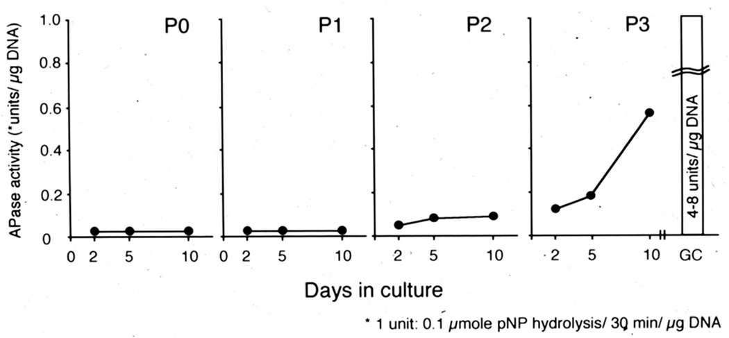
Figure 2.
Hypertrophic phenotype is induced in articular chondrocytes over time. Articular chondrocytes were isolated from femoral and tibial articular cartilage of 4-weeks old New Zealand rabbits and cultured at high density (25,000 cells/cm2) on the collagen-coated dish. Ten days after the plating, the cells were detached with trypsin/collagenase digestion and re-plated under the same condition. The passage was repeated three times (P1-P3). Primary (P0) and P1-P3 passaged cultures were subjected to measurement of alkaline phosphatase (APase) activity on Day 2, 5 and 10. The articular chondrocytes did not show APase activity in P0 and P1 cultures, increased it in P2 cultures and strongly induced in P3 cultures.

