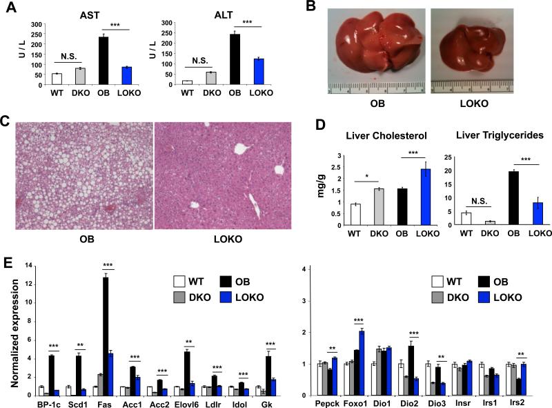
Figure 2. LOKO livers are rescued from hepatic steatosis.
(A) Markers of hepatocellular necrosis (AST, ALT) were measured by colorimetric assay in WT, DKO, OB, and LOKO mice (N = 6-8 mice per group). (B) LOKO livers are smaller and lack significant steatosis by gross examination. (C) Representative H&E sections from OB and LOKO mice are shown. Magnification = 100×. (D) Hepatic lipids were extracted and quantified from WT, DKO, OB, and LOKO mice (N = 5-6 mice per group). (E) Hepatic gene expression was measured in WT, DKO, OB, and LOKO mice (N = 5-9 mice per group) at 24 weeks of age. Results are normalized first to expression of the housekeeping gene 36B4 and shown as fold induction over WT animals. Values are means +/− SEM. Statistical analysis by 1-way ANOVA with Bonferroni post-tests: *, P < 0.05; **, P < 0.01; ***, P < 0.001, N.S. not significant.

