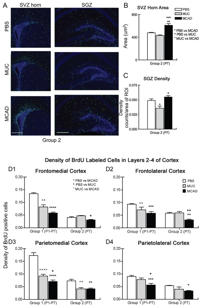
Figure 2.
A, B, and C, MCAD-treated mice have increased BrdU-positive cell proliferation in neurogenic niches (group 2). A. Immunohistochemically labeled BrdU-positive nuclei (green) in representative sections from treatment groups show increased cell proliferation in both the SVZ anterior horn and DG that includes the SGZ born neurons. B. MCAD significantly increased neurogenic niche proliferative activity at P7 in sharp contrast to MUC. C. Image-J quantified counts of BrdU-positive cells densities indicate that MCAD significantly increased neurogenic niche proliferative activity at P7 again in sharp contrast to MUC. *P < 0.05; **P < 0.01, ***P < 0.001; scale bar = 200 μm D. Decreased density of BrdU-positive cells in cortex (group 1 and 2). D1 and 2. Frontal cortex: a significant decrease in BrdU-positive cell densities in MCAD versus MUC treated groups detected in both the medial and lateral frontal cortex for group 2. D3 and 4. Parietal cortex: significant decreases in BrdU-positive cell densities in MCAD versus the MUC group in both the medial and lateral parietal lateral cortex in group 1. Although sample sizes were lower in group 2 the results complement group 1 data. *P < 0.05; **P < 0.01; ***P < 0.001; ****P < 0.0001.

