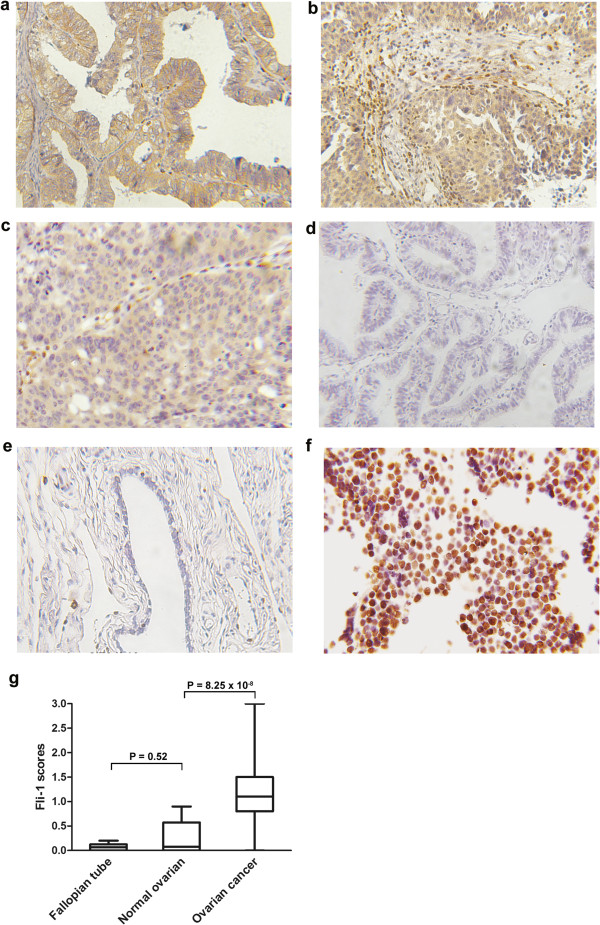
Figure 1.

Fli-1 is highly expressed in ovarian cancer. (a, b, c). Representation of images from immunohistochemical stains Fli-1 in tumors from three cases of ovarian cancer. (d) Expression of Fli-1 in fallopian tube was negative. (e) Expression of Fli-1 in normal ovarian tissue was negative. Original magnification × 200. (f) Fli-1 were positive in nuclear in PNET tissues. PNET: Primitive neuroectodermal tumor. (g) Fli-1 expression scores are shown as box plots, in ovarian cancer, fallopian tube and normal ovarian tissue. with the horizontal lines representing the median; the bottom and top of the boxes representing the 25th and 75th percentiles, respectively; and the vertical bars representing the range of data.
