
Fig. 2.
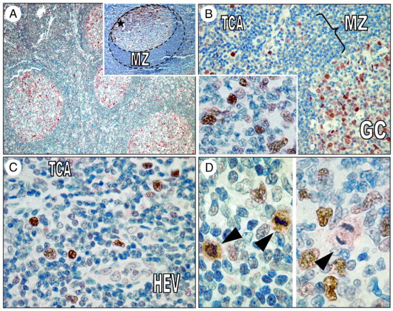
A, ser194p-FADD is detected predominantly in germinal center B cells in reactive lymph nodes and less frequently in the interfollicular areas. Note the accentuation of expression in the outer limits of germinal centers and the polarization toward the dark zone (inset, asterisk). B, ser194p-FADD protein is expressed in the nuclei of many centroblasts and some centrocytes (inset) in reactive germinal centers at variable intensity. C, In the interfollicular T-areas, ser194p-FADD expression highlights larger lymphocytes, a pattern reminiscent of Ki-67 immunostaining. D, All cells showing cytoplasmic ser194p-FADD expression were in various stages of mitosis (left and right, arrowheads). GC indicates germinal center; MZ, mantle zone; TCA, interfollicular T-cell area; HEV, high endothelial venule.