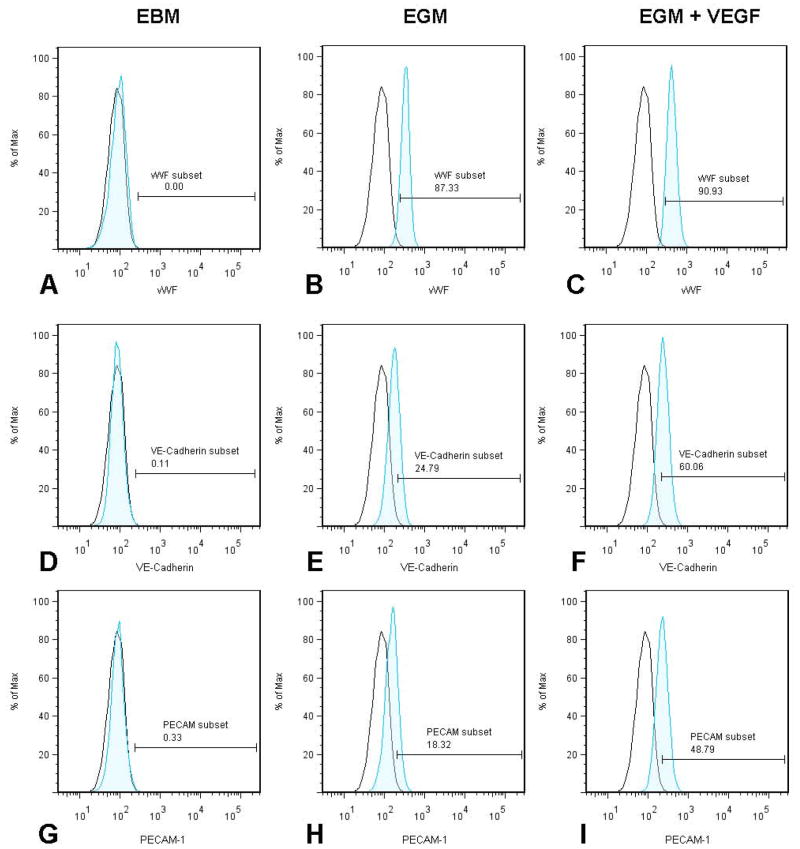
Figure 5. Immunophenotyping of differentiated cells for EC markers.
Panel 1 represents the cells stimulated in EBM media for vWF (A), VE-Cadherin (D) and PECAM-1 (G). Panel 2 represents the cells stimulated in EGM media for vWF (B), VE-Cadherin (E) and PECAM-1 (H). Panel 3 represents the cells stimulated in EGM + VEGF media for vWF (C), VE-Cadherin (F) and PECAM-1 (I). The unshaded peak shows the profile of the isotype control.

