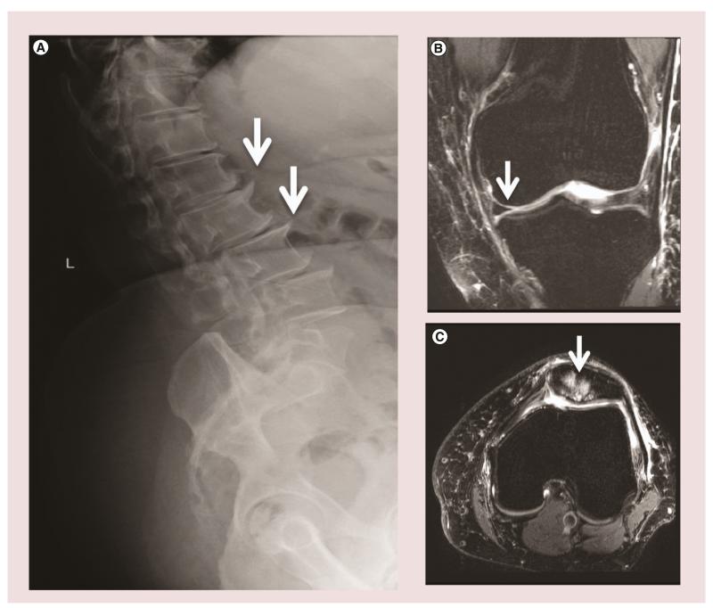
Figure 2. Radiographic features of tissue damage in osteoarthritis.
(A) Example of osteophytes (white arrows) shown in the anterior lumbar vertebral bodies. (B) MRI with T2-weighted sequences demonstrating cartilage loss (white arrow) in patient with osteoarthritis. (C) MRI with T2-weighted sequences demonstrating bone marrow lesions localized to the knee patella (white arrow) in a patient with osteoarthritis. Image acquisition paradigm for MRIs courtesy of Franklyn Howe (St George’s University, London, UK).

