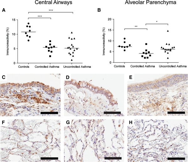
Figure 4.

Percentage area of MMP-9 (% positively stained area) in bronchial (A) and transbronchial biopsies (B) in controls and patients with controlled and uncontrolled asthma. Representative micrographs of staining of MMP-9 (brown) from controls (C, F) and patients with controlled asthma (D, G) and uncontrolled asthma (E, H) in bronchial (C-E) and transbronchial (F-H) biopsies. Scale bars: C-E and G = 100 μm, F and H = 200 μm.
