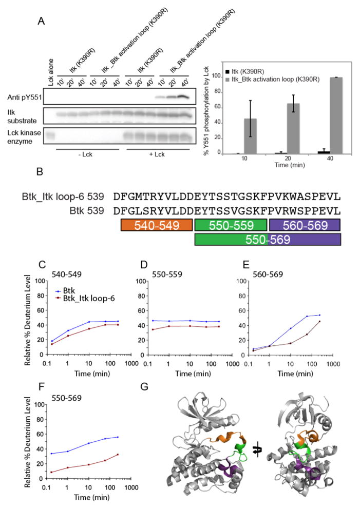
Fig. 5. Tyr511 of Itk is more accessible for phosphorylation by Lck when in the context of the Itk_Btk activation loop.
(A) Kinase-deficient (K390R) mutants of Itk or the hyperactive Itk_Btk activation loop proteins were subjected to phosphorylation by Lck in an in vitro kinase assay. Phosphorylation of Itk Tyr511 was monitored at three time points (10, 20, and 40 min) by Western blotting analysis with an anti-pY511 antibody. The Western blot is representative of three independent experiments. Densitometric data from all three experiments were quantified and are presented in the bar graph on the right. To normalize for exposure times between Western blots from different experiments, we set the abundance of pY551 in the Itk_Btk activation loop (K390R) mutant after 40 min of stimulation to 100% in each independent experiment. The abundances of the phosphorylated Itk (K390R) protein (all three time points) and Itk_Btk activation loop (K390R) protein (at the 10- and 20-min time points) are shown relative to that for the Itk_Btk activation loop (K390R) protein at the 40-min time point. Data summarized from three independent experiments with error bars representing SD from the mean. (B) Pepsin digestions of the WT Btk and Btk_Itk loop-6 mutant proteins produced coincident peptides, which are indicated under the sequences that covered the entire activation segments of both proteins. (C to F) Deuterium exchange was measured for the WT Btk and Btk_Itk loop-6 mutant proteins (see the full dataset in fig. S3) and data were plotted as the relative percentage of deuterium incorporation versus time for the peptides identified in (B) for Btk (blue diamonds) and Btk_Itk loop-6 (red squares). (G) The locations of the individual peptides, color-coded as in (B), are shown on the structure of the kinase domain of Btk, with the activation segment in the collapsed inactive conformation (PDB: 3GEN).

