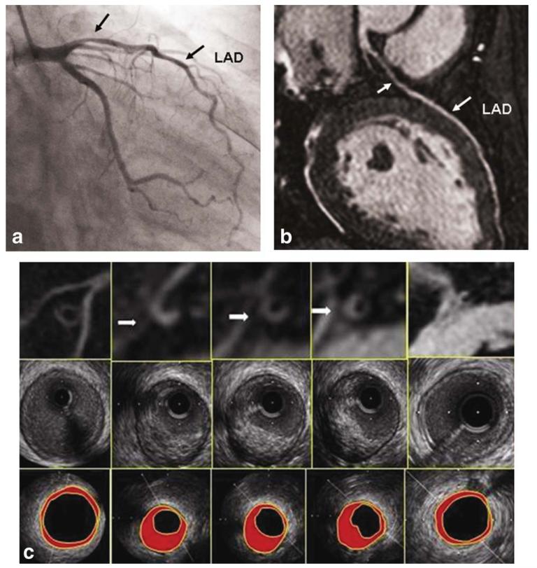
Figure 1.
A 46-year-old male participant with eccentric coronary plaque. a: LAD MRA showing moderate stenosis in the proximal coronary artery (left arrow). b: Conventional coronary artery angiography also shows moderate lumen stenosis in the same site (left arrow). c: Cross-sectional MRI coronary wall images (top row), corresponding IVUS images (middle row), and IVUS images (bottom row) from LM (right) to proximal segment of LAD (left). Plaques were found in MRI (arrows), which were correlated well with IVUS.

