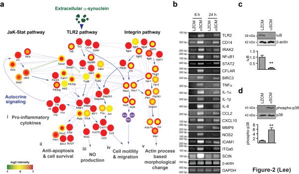
Figure 2.
Hypothetical signaling network of microglia activated by cell-released α-synuclein. (a) Hypothetical signaling network was constructed by using DEGs from αSCM-exposed microglia. Alterations of gene expression were detected at two different time points (Inner circle; 6 hours and Outer circle; 24 hours). (b) RT-PCR analysis of genes identified in the network. (c, d) IκB degradation (c) and phosphorylation of p38 MAP kinase (d) in microglia exposed to αSCM for 15 minutes (n = 4). All data were analyzed using unpaired t-test. Error bars represent ± s.e.m. **P < 0.01. “n” represents the number of independent experiments.

