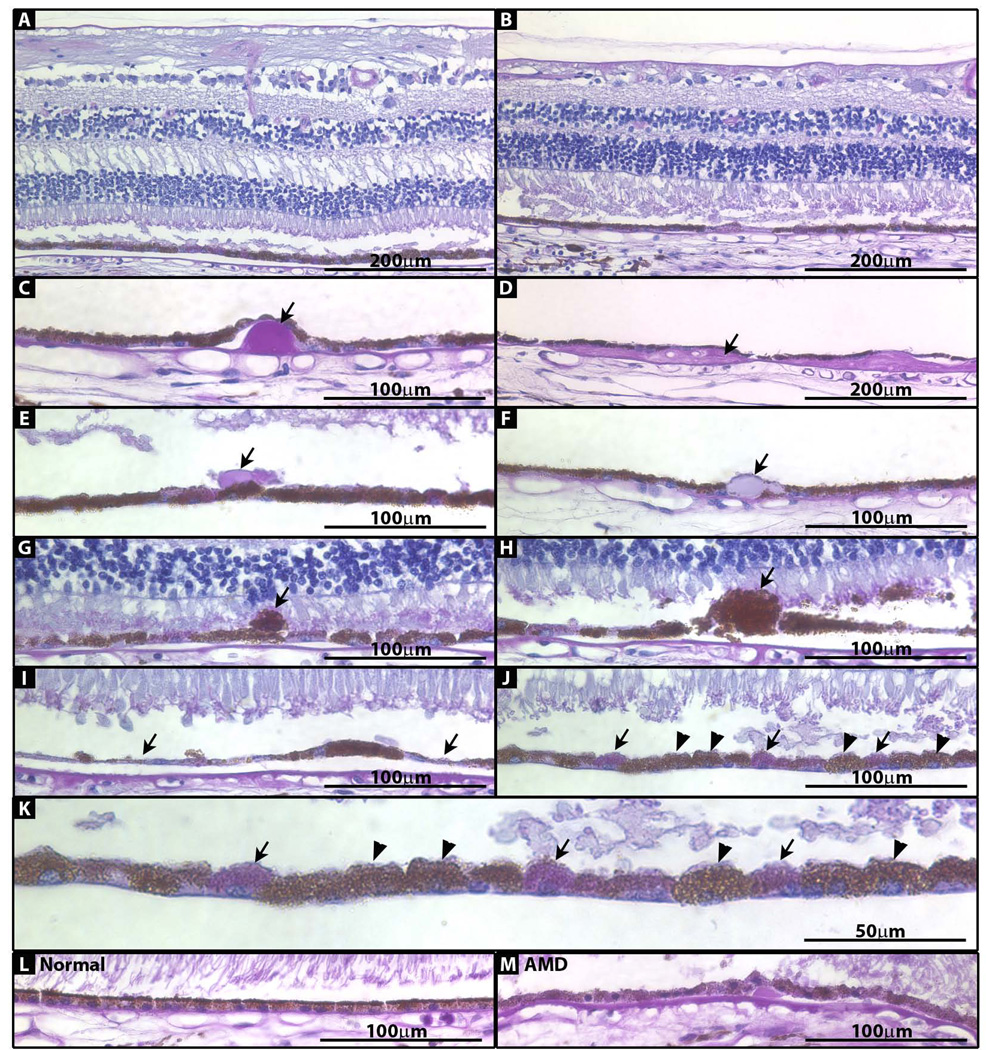
Figure 1. Photomicrographs of paraffin-embedded sections of peripheral and macular retina stained with PAS/hematoxylin.
(A) Macula. (B) Periphery. (C) Nodular druse (arrow) from periphery. (D) Diffuse druse (arrow) from periphery. (E) Subretinal drusenoid deposit (arrow) from macula. (F) RPE inclusion (arrow) from periphery. (G) Extruded RPE cell (arrow) from macula. (H) Hypertrophic RPE cell (arrow) from macula. (I) RPE thinning/atrophy (arrows) in macula. (J) Areas of pigmented (arrowhead) and depigmented (arrow) RPE cells. (K) Image in J enlarged. (L) RPE from normal control eye. (M) RPE from AMD control eye.

