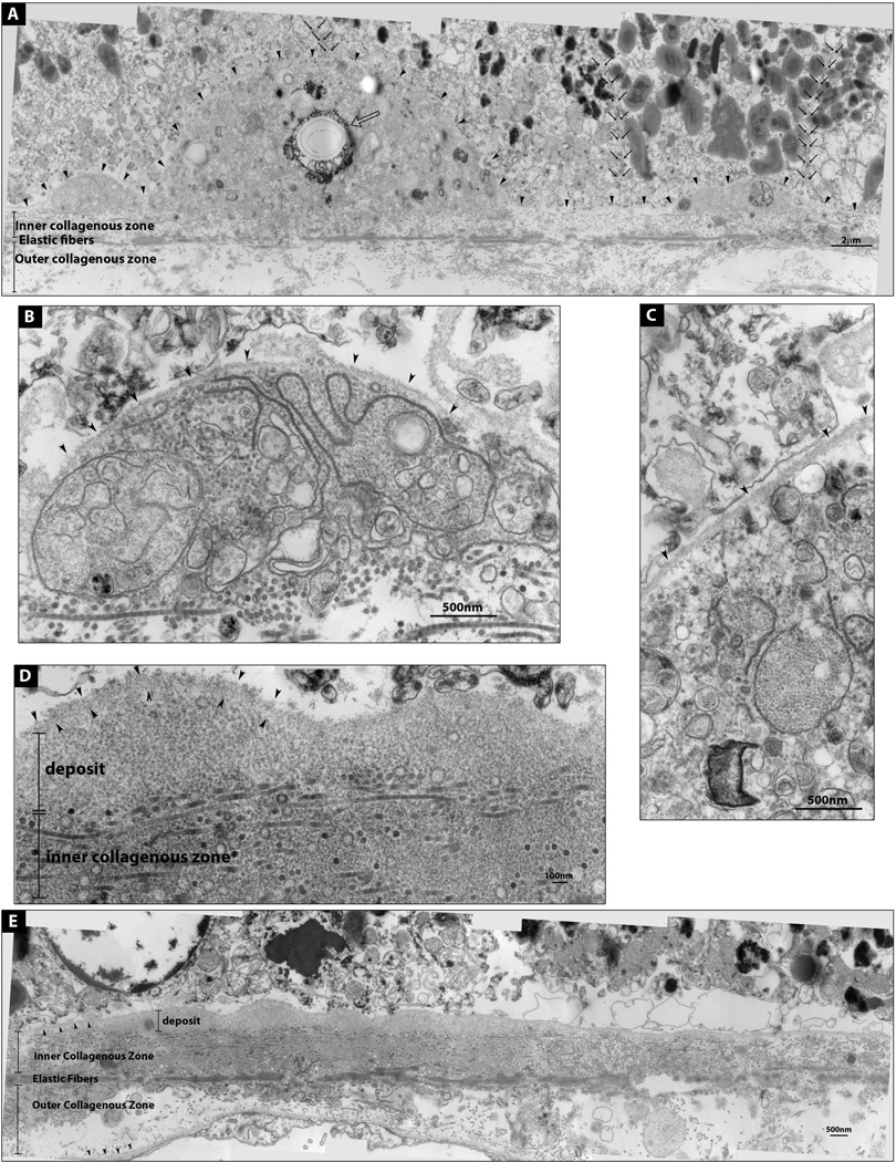
Figure 2. Electron micrographs of a druse and a basal linear deposit.
(A) A Druse flanked by two deposits. RPE basal lamina is demarcated by arrowheads. These deposits contain membranous components and a crystallized area (open arrow). Both melanosome-rich and poor RPE cells sit on top of the deposits; cell boundaries are marked by small black arrows. (B and C) Enlarged images of the deposits in A. (D) Enlarged image of E; basal linear deposit containing homogenously granular material; RPE basal lamina (arrowheads). (E) Basal linear deposit. RPE and choroidal basal laminas are marked by arrowheads.

