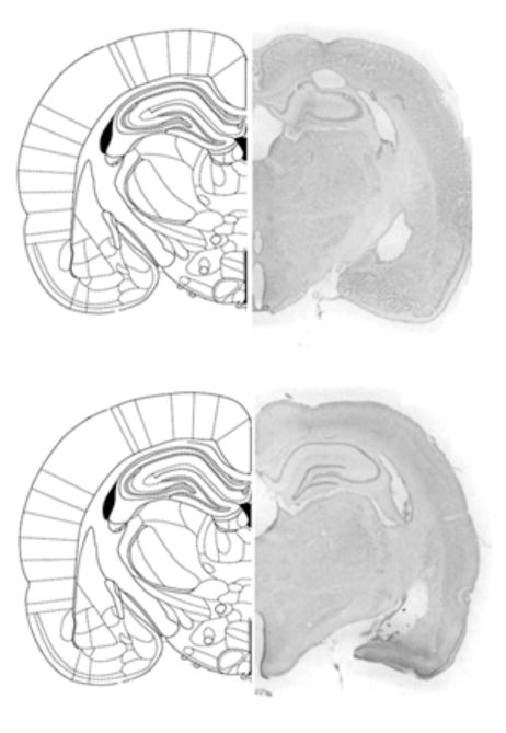
Figure 3.
Figure 3A. Histological representation of a DG lesioned rat brain and schematic drawing of corresponding coronal section (adapted from Paxinos & Watson, 1997).
Figure 3B. Histological representation of a vehicle-infused control rat brain and schematic drawing of corresponding coronal section (adapted from Paxinos & Watson, 1997).

