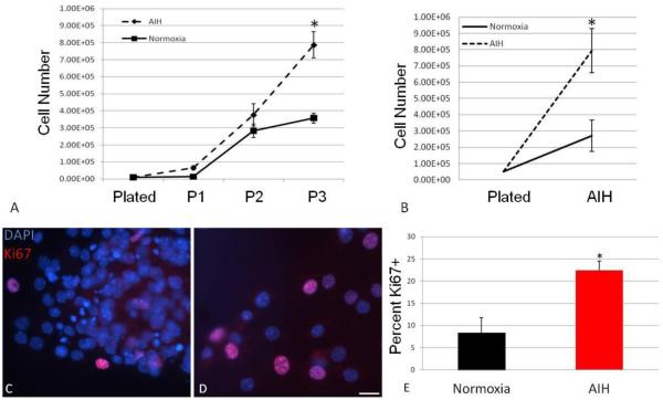
Figure. 2. In vivo AIH increases in vitro NS and MASC expansion and proliferation.
NS were serially passaged every 4-5 days and assessed for population expansion by counting single, dissociated cells with a Trypan Blue exclusion assay. Compared to normoxic (control) NS, those from AIH-treated animals exhibited progressive population expansion (A, *: P<0.001 interaction between time and treatment). In addition, three days after plating, more MASC could be detected in cultures from AIH-treated animals compared to controls (B*: P<0.001 interaction between time and treatment). In parallel wells, NS progeny were assessed for expression of the proliferation marker, Ki67. Representative images showing Ki67 (red) and DAPI immunostaining (blue) are shown in panels C (AIH-treated) and D (control) (scale bar = 25μm). Compared to control, cells from AIH-treated animals exhibited a significantly greater number of Ki67 positive cells (E, *: P<0.005).

