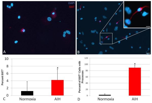
Figure 6. In vivo AIH increases neuronal differentiation in cultured MASC progeny.
MASC from control or treated animals were plated in triplicate on poly-L-ornithine coverslips and analyzed for ß-III tubulin positivity, with each replicate representing an individual animal. Compared to controls (A), cultures from AIH-treated animals (B) were observed to contain a greater number of spontaneously-formed neuroblasts prior to growth factor withdrawal. The scale bars in panel B indicate 25μm and 5 μm, respectively. Panel C shows the percentage of cells which were positive for ß-III tubulin (*, p<0.05). Panel D shows the percent of beta-3 tubulin+ cells that showed distinct processes extending from the cell body (*, p<0.0001).

