
Fig. 3.
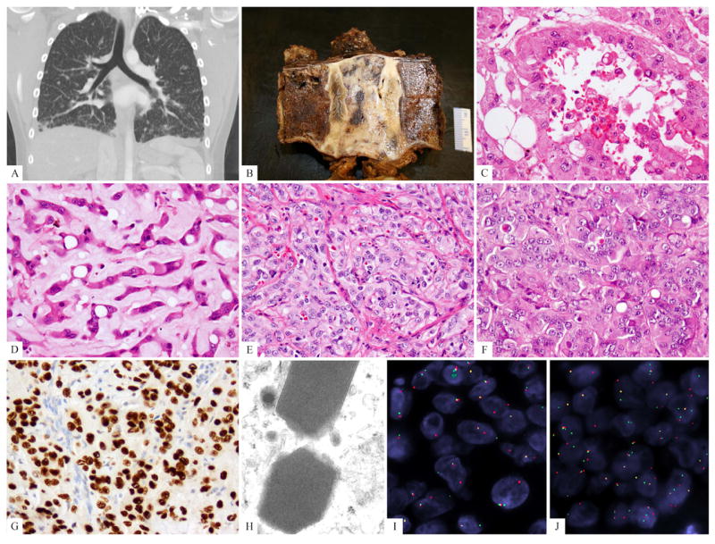
Clinical and pathologic spectrum of TFE3-rearranged EHE. (A) CT scan showing bilateral ground-glass opacities suggestive of interstitial lung disease, most predominantly in the lower lobes but diffusely present (EHE9); (B) Gross appearance of the T2 vertebral en-bloc resection showing an ill-defined white-gray lesion (EHE3). (C, D). Biphasic morphologic appearance showing a pseudo-alveolar component with abundant, densely eosinophilic cytoplasm and the other resembling classis EHE, with cord-like arrangement and myxoid stroma (EHE3); (E) foamy cytoplasm (histiocytoid), mild nuclear pleomorphism (EHE7); (F) predominantly solid and nested growth pattern, showing densely eosinophilic cytoplasm, rare vacuoles, and lack of significant intervening stroma (EHE4); (G) strong TFE3 immunostaining; (H) ultrastructural study showing distinctive rhomboid crystals with a periodicity ranging from 9.05-11.63, mean of 10.34 nm, (46,000 magnification, EHE10). (I) TFE3 and (J) YAP1 break-apart signals by FISH (EHE 7) (green, telomeric; red, centromeric).