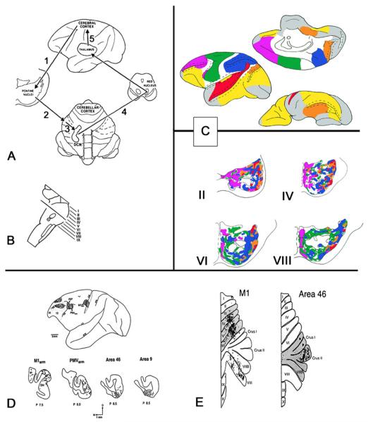
Fig. 1.
a Diagram of the cerebro-cerebellar circuit. Feedforward limb: the corticopontine pathway (1) carries associative, paralimbic, sensory, and motor information from the cerebral cortex to the neurons in the ventral pons. The axons of these pontine neurons reach the cerebellar cortex via the pontocerebellar pathway (2). Feedback limb: the cerebellar cortex is connected with the deep cerebellar nuclei (3), which course through the midbrain in the vicinity of the red nucleus to terminate in the thalamus (the cerebello-thalamic projection, 4). The thalamic projection back to cerebral cortex (5) completes the feedback circuit. b Plane of section through the pons from which the rostrocaudal levels II through VIII are taken in the schematic (c). c Composite color-coded summary diagram illustrating the distribution within selected regions of the basis pontis of projections from association and paralimbic areas shown on medial, lateral, and orbital views of the cerebral hemisphere in the prefrontal (purple), posterior parietal (blue), superior temporal (red), and parastriate and parahippocampal regions (orange) and from motor, premotor, and supplementary motor areas (green). Other cerebral areas known to project to the pons are depicted in white. Cortical areas with no pontine projections are shown in yellow (from anterograde and retrograde studies) or gray (from retrograde studies). Dashed lines in the hemisphere diagrams represent sulcal cortices. Dashed lines in the pons diagrams represent pontine nuclei; solid lines depict corticofugal fibers. d Lateral view of monkey brain (top) shows the locations of viral tracer injections in the M1 arm, PMv arm, and prefrontal cortex areas 46 and 9. The resulting retrogradely labeled neurons in the cerebellar dentate nucleus (bottom) are indicated by solid dots. e Flattened representation of cerebellum to show the folia linked with M1 motor cortex (left) and prefrontal cortex area 46 (right) using viral tracers that travel in the anterograde direction (H129 strain of HSV1) and retrograde direction (rabies virus). a–c reproduced and adapted from Schmahmann [62] and Schmahmann and Pandya [34, 35, 51]; d from Middleton and Strick [208]; e from Kelly and Strick [30]

