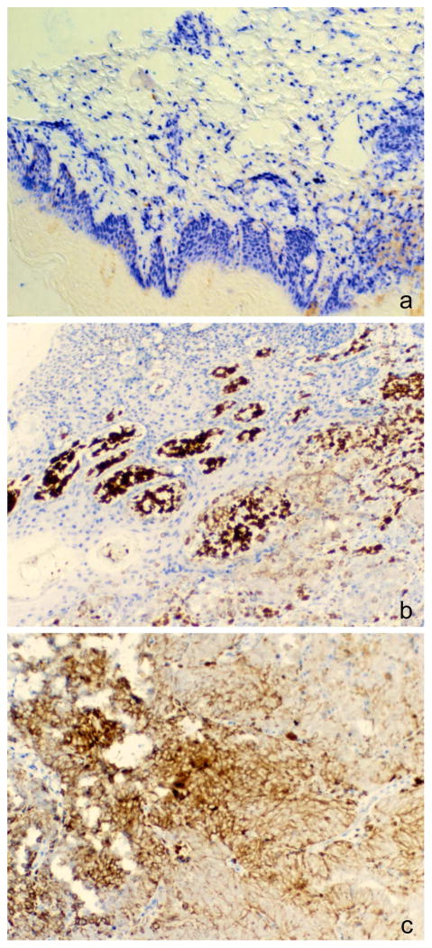
Figure 3. Reduced staining of 5-hydroxymethylcytosine in a melanoma biopsy.
Different sections of the same biopsy showing an area of normal skin (A), normal skin adjacent to a melanoma (B; tumor in lower right quadrant) and a melanoma tumor section (C) were stained with anti-5hmC antibody and AP-blue dye (Blue Alkaline Phosphatase Substrate Kit, Vector Laboratories).

