
Figure 5. Immuno-fluorescent labeling of serotonergic raphespinal projections in WT and GFAP-IκBα-dn spinal cords 10 weeks after contusive SCI.
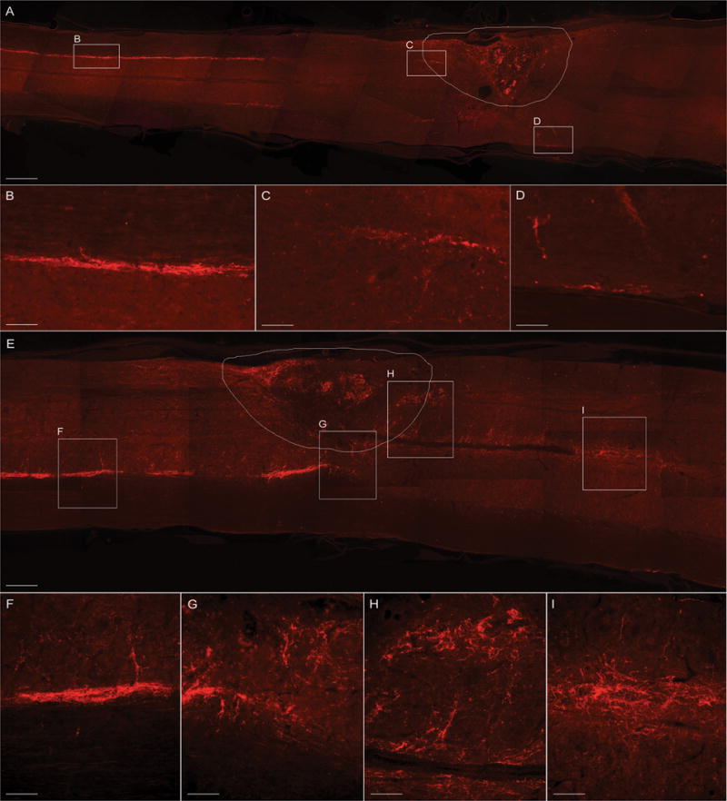
Raphespinal axons were identified by immunolabeling with an anti-5-HT antibody in longitudinal sections of the spinal cord cut along the horizontal plane. WT mice (A–D) showed minimal 5-HT labeling below the injury site, contrary to GFAP-IκBα-dn mice (E–H), who showed a significant presence of 5-HT-labeled raphespinal projections fibers throughout and below the lesion site (H, I). Convoluted and arborized fibers in GFAP-IκBα-dn mice are an anatomical indication of collateral regenerative sprouting (I). The contour in A and E delineates the lesion area. Scale bars: A, E = 500 μm; B, C, D = 50 μm; F, G, H, I: 80 μm.