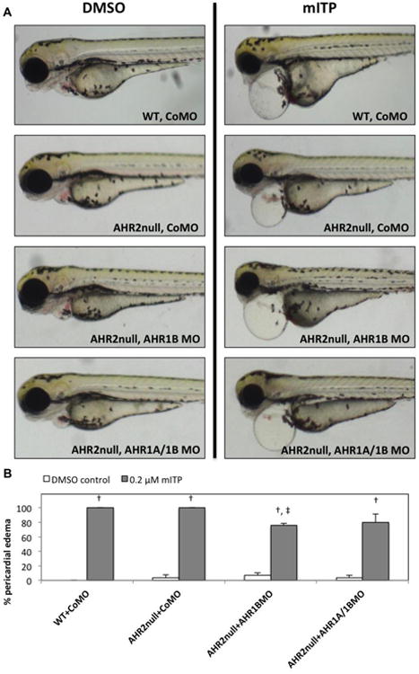
Figure 2.

mITP-induced cardiotoxicity is AHR-independent. 5D embryos (WT) were injected with CoMO, and ahr2hu3335 mutant embryos (AHR2null) were injected with either CoMO, AHR1B MO, or co-injected with AHR1A and AHR1B MOs. (A) Representative bright-field images of WT and AHR2null morphants exposed to either vehicle control (0.1% DMSO) or 0.2 μM mITP. (B) Percent PE is mean ± SE. All groups were allowed to develop until 72 hpf. Dagger (†) denotes a statistically significant increase in PE in mITP-exposed embryos relative to vehicle controls within the same group (p < 0.05), whereas double dagger (‡) denotes a statistically significant decrease in PE relative to mITP-exposed CoMO-injected WT fish (p < 0.05). N = 3 replicate vials and 9 to 10 fish per replicate.
