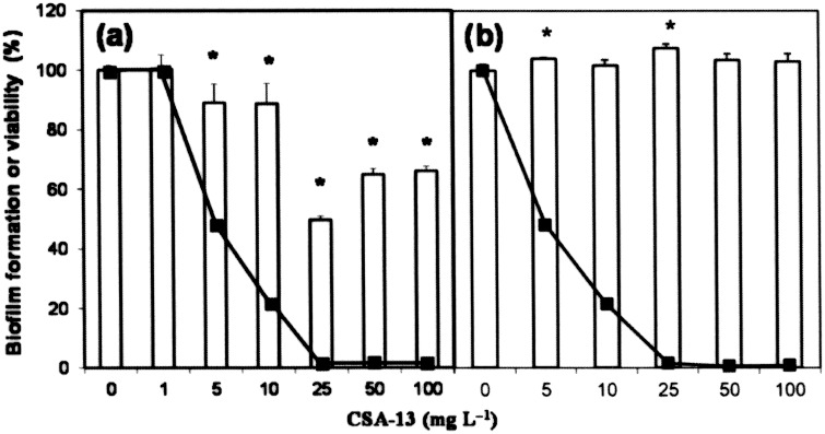
Figure 6. Disaggregation of biofilms in the presence of CSA-13.
S. pneumoniae R6 (a) and P103 (lytA::aphIII) (b) were inoculated into 200 µl of C medium in the wells of a microtiter plate (4.5×10⁶ CFU/ml). After biofilm development (6 h at 34°C), CSA-13 was added at different concentrations, and incubation allowed to proceed for 1 h at 34°C before staining with crystal violet to quantify biofilm formation (open bars). Percentage viability (solid lines) after treatment with CSA-13 was determined by plating on blood agar plates. Standard error bars are shown. Asterisks indicate a P value<0.05.

