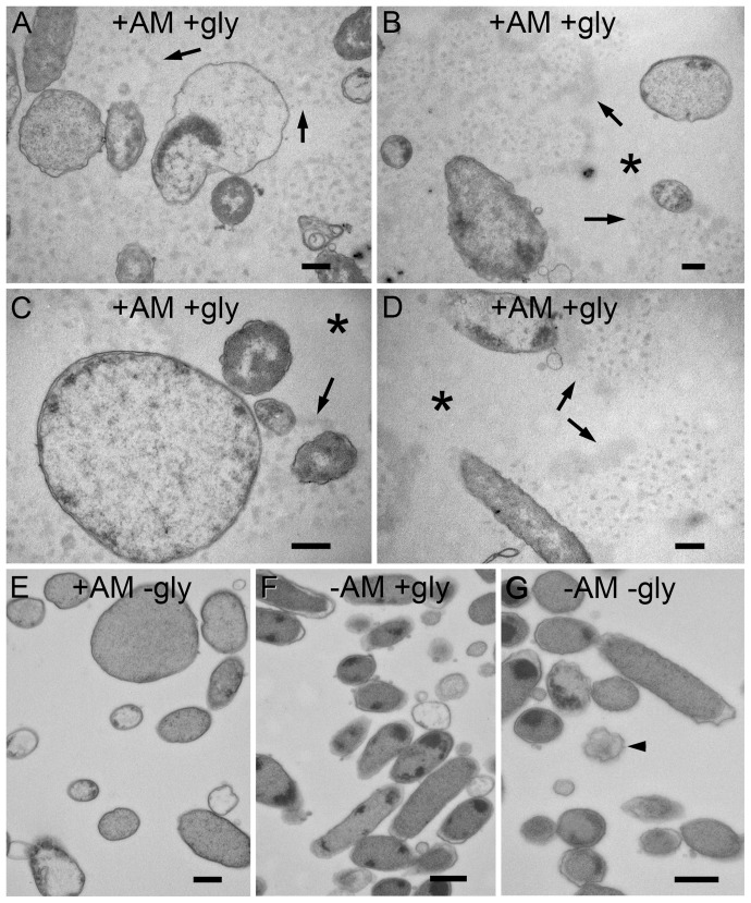
Figure 9. Ultrastructural visualization of glycogen in PittGG biofilms.
Resin-embedded thin sections of PittGG biofilms formed in the presence or absence of ampicillin (+/− Am) and treated with a glycogen stain or a no stain control (+/− gly). A–D Biofilms with ampicillin, sections stained for glycogen (periodic acid and sodium chlorite). The glycogen stain reacted with an extracellular granular substance (arrows) that was associated with biofilm bacteria. Exposure to antibiotic resulted in a bacterial size increase with some bacteria (A–C). Extracellular areas that were not in close proximity to bacteria cells (asterisk) did not contain the extracellular granular substance (B–D). E. Biofilm with antibiotic but only treated with sodium chlorite did not reveal extracellular granular substance. Large bacterial cells were present. F. Biofilm with no antibiotic, sections stained for glycogen. No extracellular granular substance was detected and bacterial cells had a normal diameter (approx. 500 nm). Some lysed cells were detected. G. Biofilm with no antibiotic, treated with sodium chlorite (no periodic acid). Bacterial cells appear normal with a few dying cells present (arrowhead). Scale bars = 500 nm.

