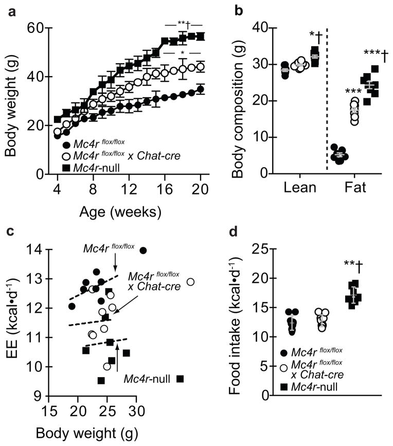
Figure 1.
Deleting melanocortin 4 receptors (Mc4rs) in pre-ganglionic cholinergic neurons in the sympathetic nervous system causes obesity due to reduced energy expenditure (EE). (a) Body weight in chow-fed mice with intact Mc4r signaling (controls; Mc4rflox/flox), selective deletion of Mc4rs in cholinergic pre-ganglionic neurons (Mc4rflox/floxx Chat-cre), or ectopic ablation of Mc4rs (Mc4r-null) (n = 10, 12, and 9, respectively). * and ** indicate p<0.05 or 0.01 versus Mc4rflox/flox mice and † indicates p<0.05 versus Mc4rflox/floxx Chat-cre mice at indicated ages using one-way ANOVA followed by Tukey’s post-hoc analyses. (b) Body composition in chow-fed 20 week-old Mc4rflox/flox, Mc4rflox/floxx Chat-cre, and Mc4r-null (n = 8, 8, and 7, respectively) littermate mice using NMR. * and *** indicate p<0.05 or 0.001 versus Mc4rflox/flox mice and † indicates p<0.05 versus Mc4rflox/floxx Chat-cre mice at indicated ages using one-way ANOVA followed by Tukey’s post-hoc analyses. (c) Energy expenditure (EE) and (d) food intake (EE) in 7- to 8-week-old chow-fed Mc4rflox/flox, Mc4rflox/flox x Chat-cre, and Mc4r-null littermate mice (n = 9, 9, and 7, respectively) over 5 d in TSE Metabolic Cages. Dashed lines in panel c denote linear regression of EE versus body weight in 7- to 8- week-old chow-fed male mice using ANCOVA. ** and † indicate in panel d indicate p<0.01 versus Mc4rflox/flox or Mc4rflox/floxx Chat-cre mice, respectively, at indicated ages using one-way ANOVA followed by Tukey’s post-hoc analyses. All mice were male and results are means ± SEM.

