
Figure 2.
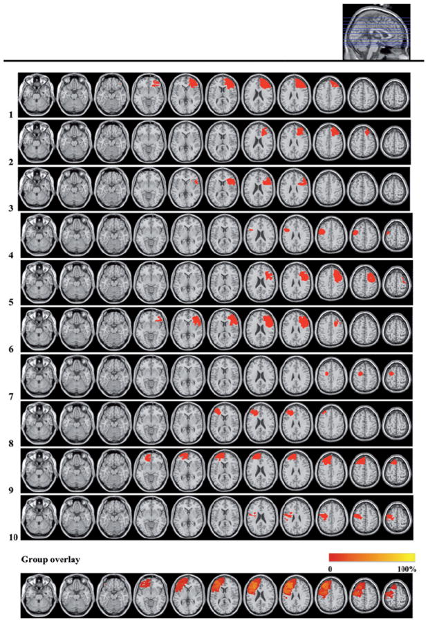
Lesion reconstructions of the LPFC group. Individual patients (1–10) and group overlay (bottom row). Seventy-one percent of the cortical lesion volume was within Brodmann Areas 6, 8, 9, 44, 45 and 46.
* Note that the group overlay presents all patients as having left hemisphere lesions to aid comparison