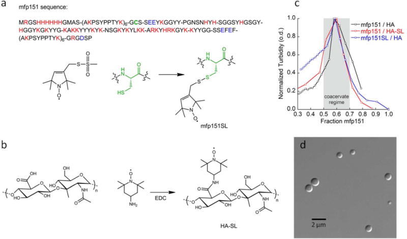
Figure 1.

(a) mfp151 sequence, where mfp1 segments (AKPSYPPTYK)6 flank the mfp5 mid-segment. Cationic residues are indicated by red and anionic residues are indicated by blue. The single cysteine is indicated in green. Mfp151 is spin labeled (mfp151SL) by covalent functionalization of the single cysteine by the spin label MTSL; (b) Hyaluronic acid (35 kDa) is spin labeled (HA-SL) by addition of 4-amino-TEMPO in the presence of EDC; (c) Turbidity of mfp151 / HA shows maximum coacervation occurs at 60 % mfp151 by mass; (d) Differential interference contrast (DIC) micrograph of mfp151 / HA complex coacervate suspension in water.
