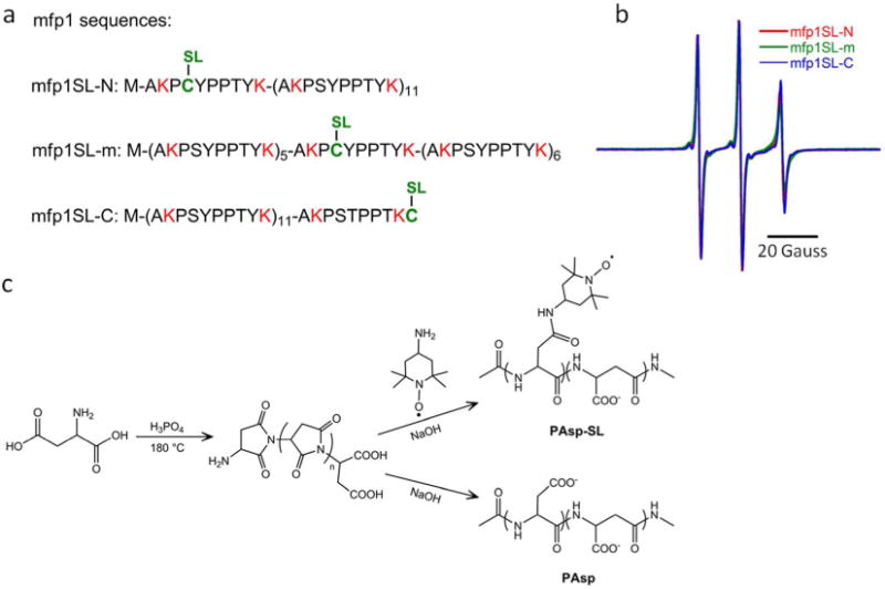
Figure 4.

(a) The mfp1 protein mutated at three select sites is then spin labeled (SL) by addition of MTSL, yielding mfp1SL-N, mfp1SL-m, and mfp1SL-C. Positively charged residues are designated in red and the single cysteine site at which the spin label is installed is indicated in green. No negatively charged residues occur in the mfp1 sequence. (b) EPR of each mfp1SL species indicates similar local environments when the polymer chains are freely dissolved in water; (c) Synthetic routes to poly(aspartic acid) (PAsp) and spin labeled poly(aspartic acid) (PAsp-SL).
