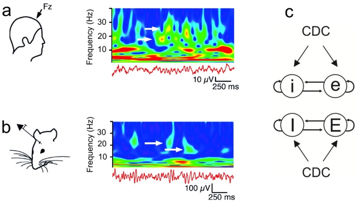
Figure 1. Distinct oscillation frequencies appearing intermittently in time, and schematic representation of model networks.
a. Wavelet of EEG recordings in the frontal zone of the human brain, showing two interspersed, non-harmonic frequencies (17.7±0.8 Hz and 22.9±0.8 Hz; white arrows) in the beta range. Color indicates power of oscillations. When one of the frequencies has high power, the other oscillation frequency is absent or has low power. Adapted from [26] (Fig. 12 therein). b. Intracranial field recordings in the medial prefrontal cortex of awake rats show similar dynamics. The two main frequencies are 15.8±0.3 Hz and 22±1.7 Hz (white arrows). Adapted from [26] (Fig. 12 therein). c. Schematic representation of the two model networks, each consisting of a population of excitatory cells (e, E) and a population of inhibitory cells (i, I). The network generating slow oscillations is labelled with lower case letters, and the network producing fast oscillations is labelled with upper case letters. In each network, the inhibitory cells projected among each other and to the excitatory cells. Likewise, the excitatory cells projected among each other and to the inhibitory cells. In addition, both cell types received external input in the form of a constant depolarizing current (CDC). Furthermore, the cells of one network projected to the cells of the other network (not shown). The different inter-network connectivity schemes studied are shown in Fig. 2.

