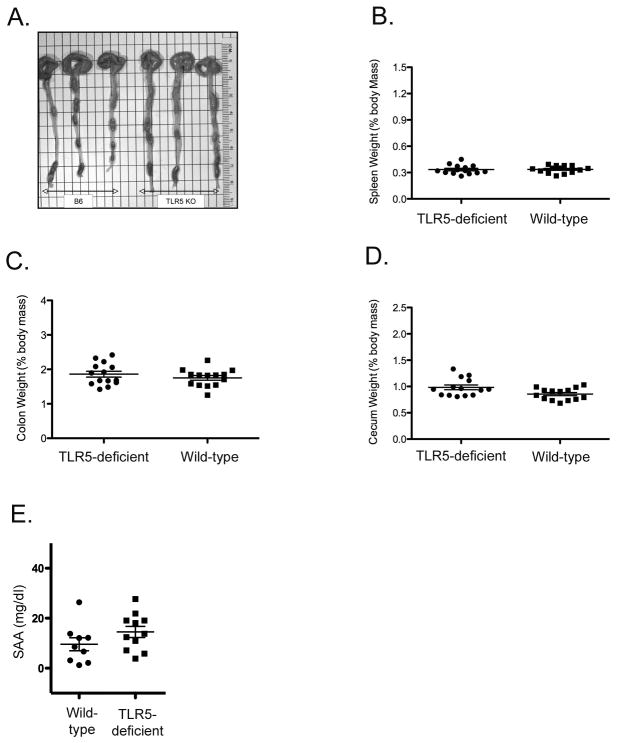
Figure 1. Lack of basal inflammatory phenotype in TLR5 Deficient mice.
Age-matched wild type and TLR5 deficient mice were euthanized at 10–12 weeks and examined for evidence of basal inflammation. (A) Photograph showing intestinal length from wild-type and TLR5-deficient mice. (B, C, D) Spleen, colon and cecum weight of wild-type and TLR5-deficient mice. Graphs show mean organ weight +/− SEM. (E) Serum Amlyoid A (SAA) levels for male wild-type and TLR5-deficient mice were determined by ELISA. Graph shows mean SAA levels +/− SEM for 9–11 mice per group. No significant difference in SAA levels was found when comparing wild-type and TLR5-deficient mice by unpaired t-test (p=0.1627).

