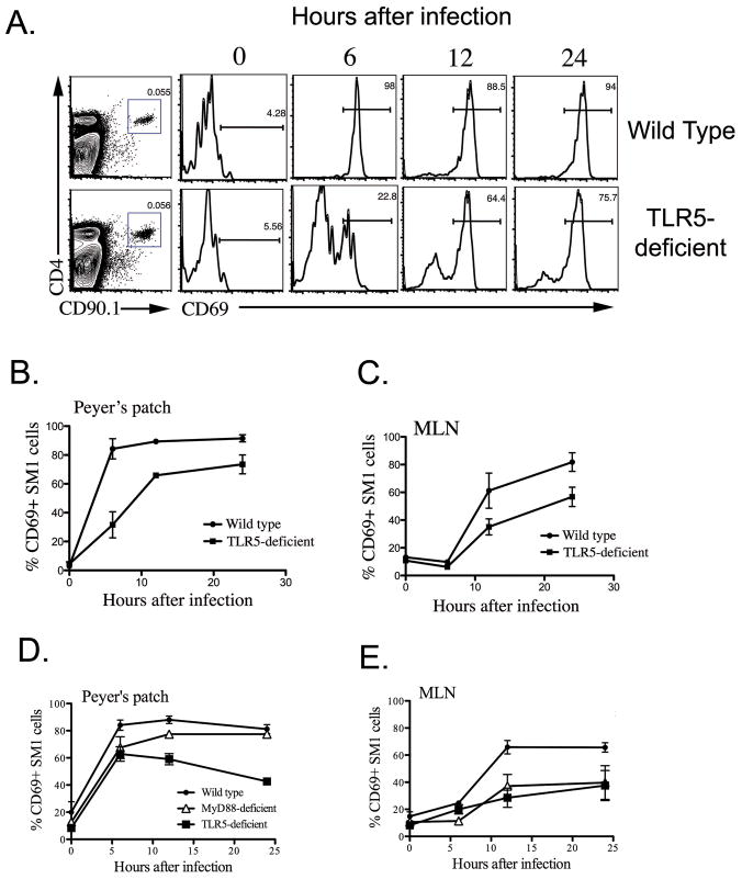
Figure 4. Deficiency in early flagellin-specific CD4 T cell activation after oral infection of TLR5-deficient mice with a flagellated pathogen.
Wild-type and TLR5-deficient mice were adoptively transferred with 800,000 SM1 T cells and infected orally with 5x10⁹ virulent Salmonella (SL1344) the following day. At various time points after infection, Peyer’s patches and mesenteric lymph nodes were harvested from mice and SM1 T cells identified using antibodies specific for CD4 and CD90.1. The early activation of SM1 T cells was examined by determining the percentage of cells expressing CD69 following infection. (A) Representative FACS plots showing the detection of SM1 T cells in Peyer’s patches (Left plot) and subsequent activation of SM1 T cells by monitoring CD69 expression after gating on SM1 cells. (B–D) Graphs show mean percentage +/− SD of CD69+ SM1 T cells in Peyer’s patch and Mesenteric lymph node after infection.

