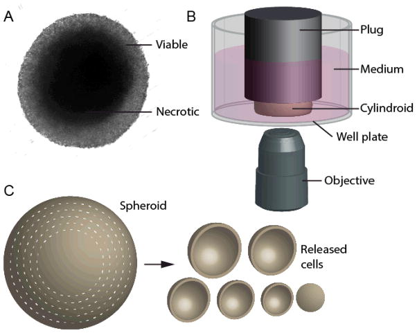
Figure 1. In vitro tumor-mimicking techniques: cylindroids and spheroid dissociation.
A) Image of a multicellular tumor spheroid, with a transparent viable periphery and an opaque necrotic core. B) Tumor cylindroids are formed by constraining spheroids between the bottom of a well plate and a plug attached to the well-plate lid. Plugs are spaced 150 μm above the bottom of the plate. The wells are filled medium and interior regions of cylindroids can be observed from the underside by microscopy. C) The metabolic content of cells as a function of radius is determined by successive rounds of dissociation that isolated cells from concentric shells.

