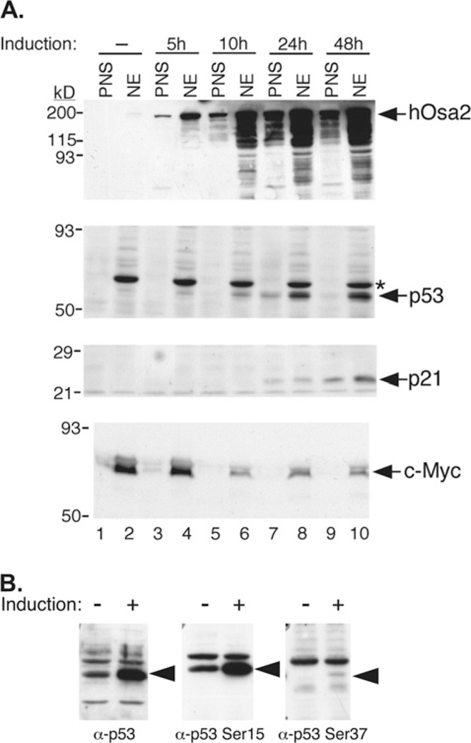
Figure 4. Changes in p53, p21 and c-Myc protein levels after induction of hOsa2.
(A) 16eC3 cells were treated with Dox for the indicated hours and then were harvested and fractionated into NE (nuclear) and PNS fractions. Proteins were separated by SDS/PAGE and immunoblotted with antibodies against hOsa2, p53, p21, or c-Myc. Molecular size markers are indicated at left. (B) Lysates of 16eC3 cells induced for 24 h (+) were separated by SDS/PAGE and probed for total p53, p53 phospho-Serine 15, and p53 phospho-Serine 37. *Non-specific cross-reacting protein.

