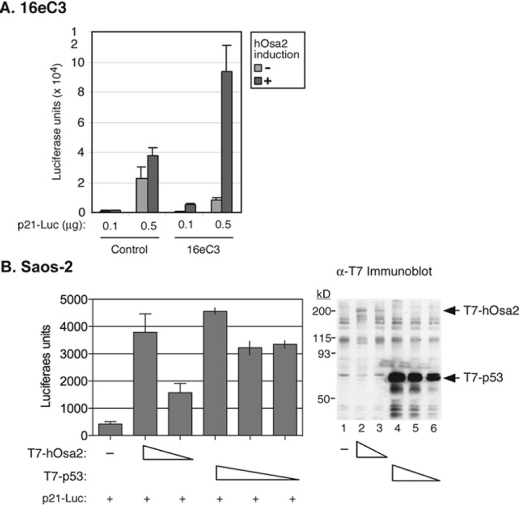
Figure 6. Activation of the p21 promoter upon co-expression of hOsa2.
(A) 16eC3 and control cells were transiently transfected by the calcium phosphate precipitation method with a p21 promoter-driven luciferase reporter plasmid with the indicated amounts of DNA. The expression of hOsa2 was induced (+) the following day by removal of Dox. The cells were harvested and luciferase activity measured after 24 h. (B) Saos-2 cells were transiently transfected with p21 promoter-reporter plasmid (0.1 µg), a plasmid expressing T7-hOsa2(16e) (1.5 and 1 µg) or a plasmid expressing T7-p53 (1, 0.5 and 0.1 µg) using the Lipofectamine™ Plus reagent. The cells were then harvested and luciferase activity measured after 24 h. The experiment was performed in triplicate (lanes 1–3) and in duplicate (lanes 4–6) and the average or the spread of the mean is shown. An immunoblot of the transfected cell lysates with anti-T7 antibody is shown in the right-hand panel.

