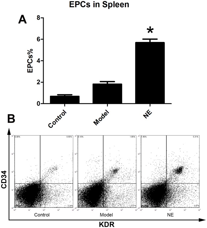
Figure 3. NE increased EPCs in spleen in mice bearing limb ischemia.
Cells from splenic tissue homogenates were lysed and analyzed with flow cytometry. EPCs in spleen were defined as CD34+/KDR+ cells (B). Proportion of EPCs in spleen was increased from 1.94±0.39% to 4.89±0.36% after intraperitoneal injection of NE in mice with limb ischemia (A, * P<0.05 compared with model group). Representative flow cytometric analysis of EPCs (CD34+/Flk-1+cells) were showed in part B.

