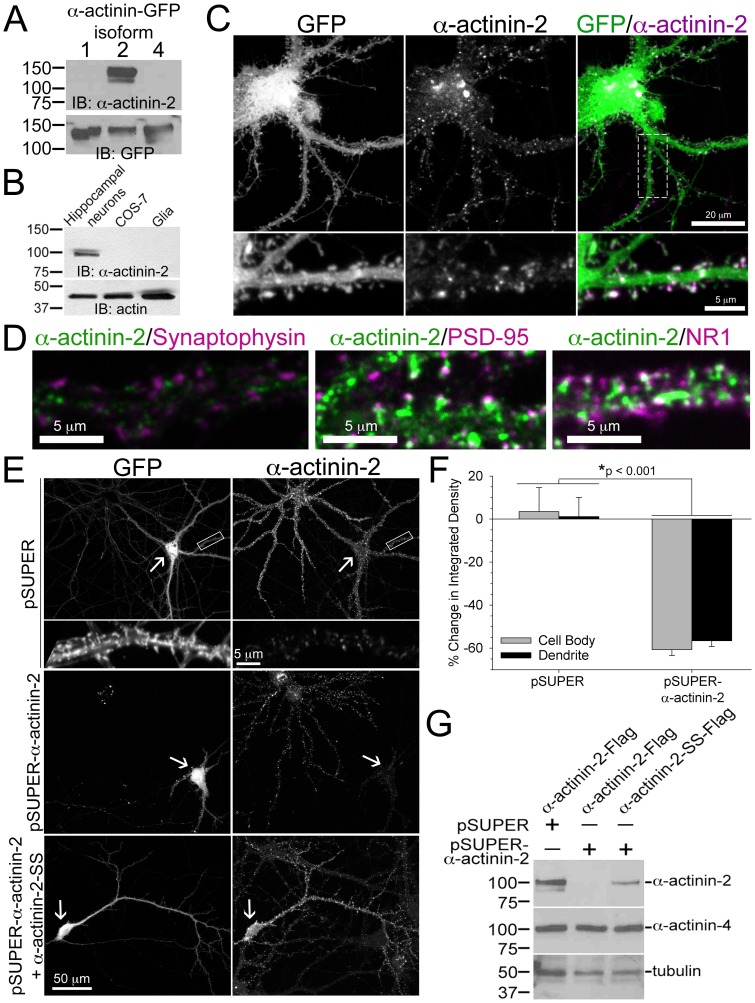
Figure 1. α-actinin-2 localizes to post-synaptic sites in dendritic spines on hippocampal neurons.
A) An anti-α-actinin antibody (ab68167) recognizes α-actinin-2 and not α-actinin-1 or α-actinin-4. CHO-K1 cells were transfected with human α-actinin-1-GFP, α-actinin-2-GFP, or α-actinin-4-GFP. Cells were lysed and immunoblotted for α-actinin-2 and GFP. B) α-Actinin-2 is enriched in hippocampal neurons but not in glia cells or COS-7 cells, which lacks α-actinin-2. Cells were lysed and immunoblotted for α-actinin-2. Actin is the loading control. C) α-Actinin-2 localizes to dendritic spines. Hippocampal neurons were transfected at DIV 17 with GFP (green), and fixed, and immunostained for endogenous α-actinin-2 (magenta) at DIV 21. D) α-Actinin-2 co-localizes with post-synaptic markers, but not with a pre-synaptic marker. Hippocampal neurons were fixed at DIV 16 or 21 and immunostained for endogenous α-actinin-2 (green) and either endogenous synaptophysin, PSD-95, or the NR1 subunit of the NMDA receptor (magenta). E–G) The siRNA is specific for α-actinin-2. Hippocampal neurons were co-transfected at DIV 17 with GFP and either a control empty vector (pSUPER), or a vector containing siRNA against α-actinin-2 (pSUPER-α-actinin-2), or the α-actinin-2 siRNA-containing vector plus a α-actinin-2 vector conferring resistance to RNAi (pSUPER-α-actinin-2+ α-actinin-2-SS). The cells were fixed at DIV 21 and immunostained for endogenous α-actinin-2. Arrows point to the neurons co-expressing GFP and its immunostaining for α-actinin-2. For each condition (55 control cells and 46 α-actinin-2 knockdown cells), the integrated density of the cell body and dendrites were measured from the transfected neuron and adjacent untransfected neuron of the same image and the percent change was plotted, F. Error bars represent SEM. p-values were derived using the paired t-test. G) CHO-K1 cells were co-transfected with GFP, pSUPER or pSUPER-α-actinin-2, plus either α-actinin-2-Flag or α-actinin-2-SS-Flag. Transfection efficiency was close to 100% as >95% of the cells in each condition exhibited GFP fluorescence (data not shown). Cells were lysed 72 hours after transfection and immunoblotted for α-actinin-2 and α-actinin-4. Tubulin is the loading control.

