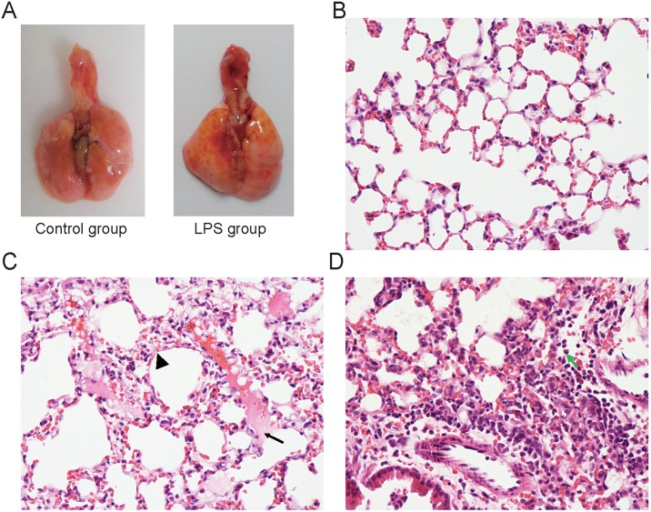
Figure 1. H&E staining shows pathological alterations that are characteristic of acute lung injury at 6 h after LPS instillation.
A: The gross appearance showed yellow FITC-Dextran on the pulmonary surface after i.n. instillation with 10 mg of FITC-Dextran/kg body weight (b.w.) for 1 hour. The lungs of the LPS model group showed red dots and swelling. B: Representative normal lung histology. C: Lung edema (arrow) and alveolar wall thickening (arrow head) in the ALI mice. D: Infiltration of many inflammatory cells (arrow shows neutrophils) in the ALI mice induced with i.n. instillation with 0.5 mg of LPS/kg b.w. for 6 h (n = 4). B, C, D the magnification is 400X.

