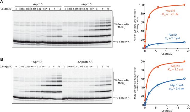
Figure 4. Activator Stimulation of Ubc4 Depends on Apc10.
(A) APC/C-Apc10Δ (~1 nM) reactions were performed as in Figure 2A with 35S-labeled securin N-terminal fragment (residues 1-110) translated in reticulocyte lysate and purified. Purified Cdh1, also produced by translation in vitro, was added to all reactions. Recombinant Apc10 was added to a final concentration of 20 μM to half the reactions as indicated, and purified Ubc4 was titrated into the reactions. Data were analyzed using Prism and fit using Michaelis-Menten parameters to determine the half-maximal E2 concentration, or apparent Km. Results are representative of two independent experiments.
(B) APC/C reactions were performed with soluble securin fragment as in (A), using immunopurified wild-type APC/C (left panel) or APC/C carrying the Apc10-4A mutant subunit (right panel). Results are representative of two independent experiments. APC/C-Apc10-4A activity is more robust than the activity of APC/C lacking Apc10 (A) because deletion of Apc10 causes a nonspecific loss of activity [15].

