
Figure 1.
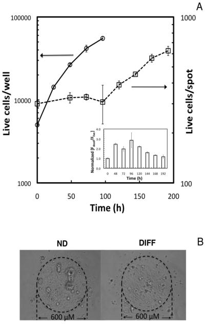
Comparison of ReNcell VM cell expansion in the 3D cellular microarray platform with expansion in conventional 2D culture platforms. (A) Cells seeded at 160 cells/mm2 in a 96-well microtiter plate (5000 cells/well; 2.5 × 10⁴ cells/ml medium) grow exponentially with doubling times of 22 h, and no lag phase observed (open circles, right axis). Cells seeded in microcultures in alginate spots at 5000 cells/mm3 (5.8 × 10⁴ cells/ml medium) demonstrate a 4-day lag, followed by a growth phase with slower proliferation (tD = 84 h) (open squares, left axis). The inset shows the changes in ReNcell VM viability during 3D culture, measured indirectly by following the ratio of average red-to-green fluorescence intensity with time, normalized to the first measurement taken after printing (typically 4 h after cell detachment). The data indicate a decrease in viability during the lag phase. Data represent the mean ± SEM. (B) Bright field images of the alginate microcultures (300 cells/spot; 5 × 10⁶ cells/ml) after cell expansion on-chip using expansion medium left panel), and cells similarly expanded and subsequently differentiated for 6 days in differentiation medium (right panel). ND refers to non-differentiated cells, DIFF refers to differentiated cells.