
Fig. 6. Effect of the CM combination on HER3 and EGFR signaling and tumor proliferation, apoptosis and angiogenesis in vivo.
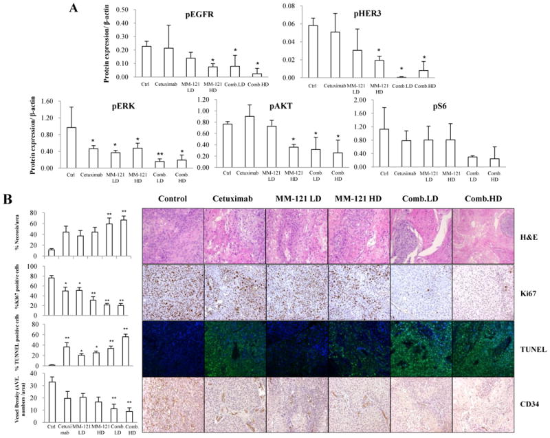
A, the CM combination inhibited HER3 and EGFR signaling in vivo. Fresh tumor tissues were collected and stored at -80°C. Three representative tissue lysates from each group were prepared for Western blot analysis. ImageJ software was used for Western blot quantification. All data are expressed as mean ± SD from 3 tissue samples. B, the CM combination significantly increased necrosis, inhibited proliferation and angiogenesis and induced apoptosis in vivo. Data show the representative tumors with hematoxylin and eosin (H&E) staining, Ki67, TUNEL and CD34 staining (Magnification: 200×). Tissue slides were observed by at least two independent personnel. The percentage of necrotic areas, percentage of positive Ki67 and TUNEL staining and CD34 positive signals were counted from five randomly selected areas in each slide at 100× magnification. For TUNEL assay, green fluorescence indicates positive cells. Error bars are mean ± SE of 7 mice from each group. (* indicates p < 0.05 and ** indicates p < 0.01 v.s. Ctrl).