
Figure 4. FOSL1, a target of BET bromodomain inhibitor, regulates growth of PDAC cells in three-dimensional collagen.
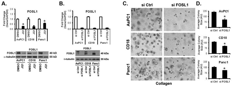
A. PDAC cells were grown in three-dimensional type I collagen gels in the presence of DMSO (vehicle control) or JQ1 (0.5 μM) for 48 hours. The effect on FOSL1 expression was analyzed by qRT-PCR using GAPDH as normalization control and by Western blotting using α-tubulin as loading control. B–D. PDAC cells were transfected with control siRNA (si Ctrl) or FOSL1-specific siRNAs (si FOSL1), allowed to recover for 48 hours, and then plated in collagen gels. The specific knockdown of FOSL1 was determined by qRT-PCR using GAPDH as normalization control and by Western blotting using α-tubulin as loading control (B). The effect on colony size was examined by phase contrast microscopy (C), and size of the individual colonies measured (D).