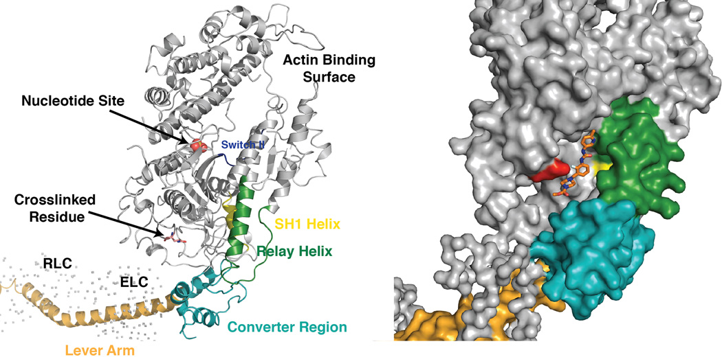
Fig. 2.
The proposed binding site for omecamtiv mecarbil to cardiac myosin S1. The ribbon diagram to the left shows the major features of the myosin S1 head. A space-filling model of the myosin structure showing the position of the identified peptide is shown to the right (the structure of the chicken skeletal S1 fragment, PDB ID 2MYS, was used as a model because the cardiac S1 structure has not been determined). The compound was manually fit into the cleft containing the identified labeled peptide. The red residue indicates the labeled amino acid serine 148.

