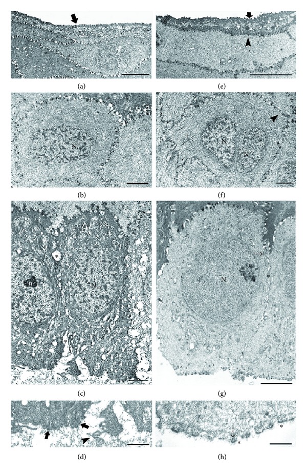
Figure 3.

(a) TEM micrograph of superficial cells from the corneal epithelium obtained with alcohol delamination on TUE. The cells are flat and show short and irregular microfolds (arrow) and dilated intercellular spaces (∗). Scale bar: 5 μm. (b) TEM micrograph of wing cells from the corneal epithelium obtained with alcohol delamination on TUE. The nucleus (N) and the cytoplasm have normal appearance; intercellular spaces are moderately dilated (∗). Scale bar: 2 μm. (c) TEM micrograph of basal cells from the corneal epithelium obtained with alcohol delamination on TUE. The cells show elliptical nuclei (N) with evident nucleoli (n), dark and vacuolated cytoplasm, and dilated intercellular spaces (∗). Scale bar: 2 μm. (d) TEM micrograph of the inferior pole of a basal cell from the corneal epithelium obtained with alcohol delamination on TUE. The cytoplasm is dense and few hemidesmosomes (arrows) are present. Note the presence of cellular blebs (arrowhead) and of an irregular granular material. Scale bar: 0.5 μm. (e) TEM micrograph of superficial cells from the corneal epithelium of TTE. The cells are flat, show regular microfolds (arrow), and are well-preserved intercellular borders (arrowhead). Scale bar: 5 μm. (f) TEM micrograph of wing cells from the corneal epithelium of TTE. Many tonofilaments (t) are evident around the apparently double nucleus (N1-N2); intercellular borders are normal, with well-evident desmosomes (arrowhead). Scale bar: 2 μm. (g) TEM micrograph of basal cells from corneal epithelium in TTE. Basal cells have round euchromatic nucleus (N), clear cytoplasm with only few vesicles, small intercellular widenings (∗), and some desmosomes (arrow). Scale bar: 2 μm. (h) TEM micrograph of the inferior pole of a basal cell from the corneal epithelium in TTE. The cytoplasm is clear and the hemidesmosomes (arrows) are numerous. ∗ = lamina lucida of the basement membrane. Scale bar: 0.5 μm.
