Figure 9.

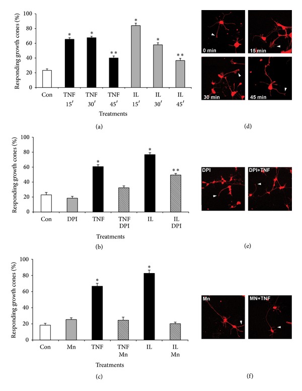
Cytokines elicit redox-dependent reorganization of actin filaments in neuronal growth cones. SC neurons grown on laminin (2 days) were incubated with 40 μM MnTBAP, 5 μM DPI or left untreated prior to addition of TNFα or IL-1β (200 ng/mL) for increasing time periods (15 min, 30 min, and 45 min). Cultures were fixed, permeabilized (Triton-X-100), and stained with rhodamine phalloidin to reveal filamentous actin. Random images were acquired (40x, confocal microscope), growth cones were scored for the presence of at least one, distinct actin filament-rich structure, and all values normalized to control conditions (% responding growth cones). (a) Both TNFα and IL-1β significantly increased the percentage of growth cones with actin-filament rich structures (*P < 0.01) at 15 min and 30 min after exposure followed by a significant decrease at 45 min after exposure (**P < 0.01 compared to t = 15 min) as opposed to control. (b) Inhibition of NOX activity with 2 μM DPI largely negated the formation of actin-filament rich structures upon exposure to TNFα (TNF-DPI) or IL-1β (IL-DPI, **P < 0.01) as opposed to TNFα only (TNFα, *P < 0.01) or IL-1β only (IL, *P < 0.01), respectively. Large actin filament-rich structures were not affected by DPI in the absence of cytokines (DPI) compared to control (Con). (c) Scavenging ROS with 10 μM MnTBAP abolished the formation of actin filament-rich structures in response to TNFα (TNFα-Mn) or IL-1β (IL-Mn) when compared to TNFα only (TNF, *P < 0.01) or IL-1β only (IL, *P < 0.01). The degree of actin filament-rich structures in the presence of MnTBAP alone (Mn) was indistinguishable from control (Con). ∗Significant difference from control and ∗∗significant difference from respective cytokine treatment at P < 0.05 by Kruskal Wallis test and Dunnett's t-test. (d–f) Cultured SC neurons were treated with pharmacological inhibitors prior to bath application of cytokines, fixed with 4% paraformaldehyde, and stained for actin filaments with rhodamine phalloidin (red signal). (d) Representative images of SC neurons with increasing exposure time to TNFα. Actin filament-rich structures in growth cones (lamellipodia) are indicated with arrowheads. (e) Incubation with DPI largely abolished the formation of actin filament-rich structures in SC neurons cultures. (f) Scavenging ROS with MnTBAP effectively negated appearance of actin filament-rich structures in growth cones of SC neurons.
