
Figure 4.
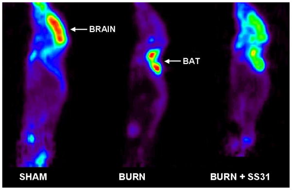
Representative 18FDG-μPET images of a sham treated mouse (Left panel), a mouse with burn injury (Middle panel) and a mouse with burn injury that was treated with SS31 (Right panel). 18F-FDG μPET imaging demonstrated intense focal uptake at sites of BAT after burn injury. Uptake in BAT was so intense that it was associated with significant reductions in uptake by all other tissues, including brain. In the mouse treated with SS-31, there was reduced 18FDG in BAT, and a partial normalization of 18FDG uptake in brain.