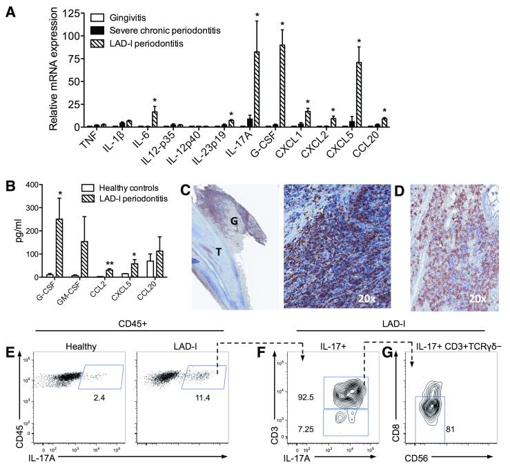
Fig. 2. IL-17 signature in LAD-I periodontitis.
(A) Quantitative real-time PCR analysis of indicated cytokine mRNA expression levels in the lesions of LAD-I periodontitis compared to those from severe chronic periodontitis (severe inflammatory bone loss) or gingivitis (gingival inflammation without associated bone loss); results were normalized to HPRT mRNA and presented as fold induction relative to gingivitis, assigned an average value of 1. Data are means ± SEM (n=4 per group). *P < 0.05 vs. both chronic periodontitis and gingivitis (one-way ANOVA). (B) The indicated cytokines/chemokines were measured in gingival crevicular fluid from healthy control subjects and LAD-I patients, using multiplex luminex assays. Data are means ± SEM (n=5 per group) *P < 0.05 and **P < 0.01; unpaired t test. (C) Immunohistochemistry for IL-17A in LAD-I gingiva [G] surrounding an extracted tooth [T] (left). Numerous IL-17A+ cells are seen throughout the lesion, shown in larger magnification (right). (D) Immunohistochemistry for CD3 in IL-17A+ cell regions. C and D are serial sections, representative of 4 patients and multiple tooth sites. (E-G) Characterization of IL-17-producing cells in LAD-I periodontitis. (E) Flow cytometry after intracellular staining for IL-17A in isolated CD45+ gingival cells, from healthy (left) or LAD-I (right) subjects, stimulated with PMA and ionomycin. Plots F and G show further characterization of IL-17+ populations in LAD-I: (F) IL-17A versus CD3 staining gated on CD45+IL-17+ cells; (G) CD56 versus CD8 staining gated on CD45+IL-17+CD3+TCRγδ− cells. Representative of two separate experiments with paired LAD-I versus healthy control comparisons.

