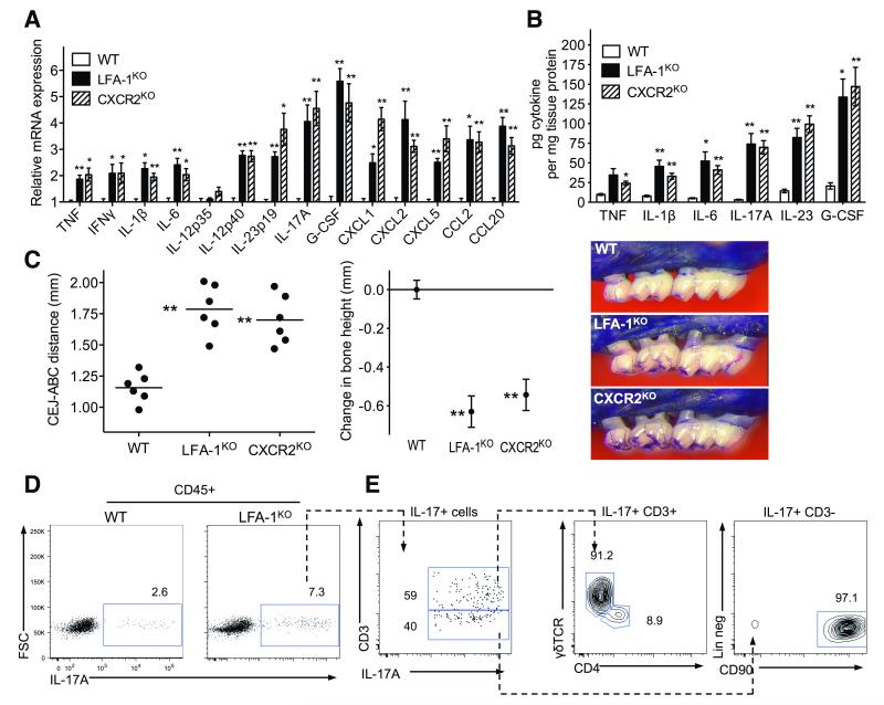
Fig. 4. Cytokine profiles in periodontitis associated with defective neutrophil adhesion and/or recruitment.
18-week-old WT control mice were compared with age-matched LFA-1KO and CXCR2KO mice for cytokine levels and bone loss. (A, B) Gingiva were dissected to assess the indicated cytokine responses at the mRNA (A) or protein (B) level. Cytokine mRNA expression levels were normalized against GAPDH mRNA and expressed as fold induction relative to the transcript levels of 18-week-old WT mice, which were assigned an average value of 1. (C) Measurement of periodontal bone heights (CEJ-ABC distance) in the indicated mouse groups (left), calculation of relative bone loss (middle), and representative images of maxillae (right). In the left panel, each symbol represents an individual mouse and small horizontal lines indicate the mean. In the middle panel, bone loss was calculated as bone height in WT control mice (0 baseline) minus bone height in experimental mice. Data are means ± SEM (n = 6 mice/group) from one of two independent experiments with similar results. *P< 0.05 and **P< 0.01 compared with WT controls (one-way ANOVA). (D, E) Flow cytometry of cell preparations isolated from mouse gingiva stimulated with PMA and ionomycin. (D) IL-17 staining in CD45+ cells from WT and LFA-1KO mice. (E) Further characterization of IL-17+ populations in LFA-1KO mice. Plots shown from left to right; staining for CD3 versus IL-17 gated on CD45+IL-17+ cells; TCRγδ versus CD4 gated on CD45+IL-17+CD3+ cells; and lineage staining (CD3−CD19−CD5−NK1.1−CD11c−CD11b−Ly6G−CD117−) versus CD90 gated on CD45+IL-17+CD3− cells. Data are representative of three independent experiments.

