
Figure 1.
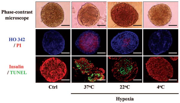
Morphological appearance of four groups of islets. Control (Ctrl), hypoxia-37°C, hypoxia-22°C and hypoxia-4°C islets were examined by phase-contrast microscopy, Hoechst33342 (blue)/propidium iodide (PI; red) staining and insulin (red)/Terminal Deoxynucleotidyl Transferase-Mediated dUTP Nick-End Labeling (TUNEL; green) staining. Scale bars: 50 μm.