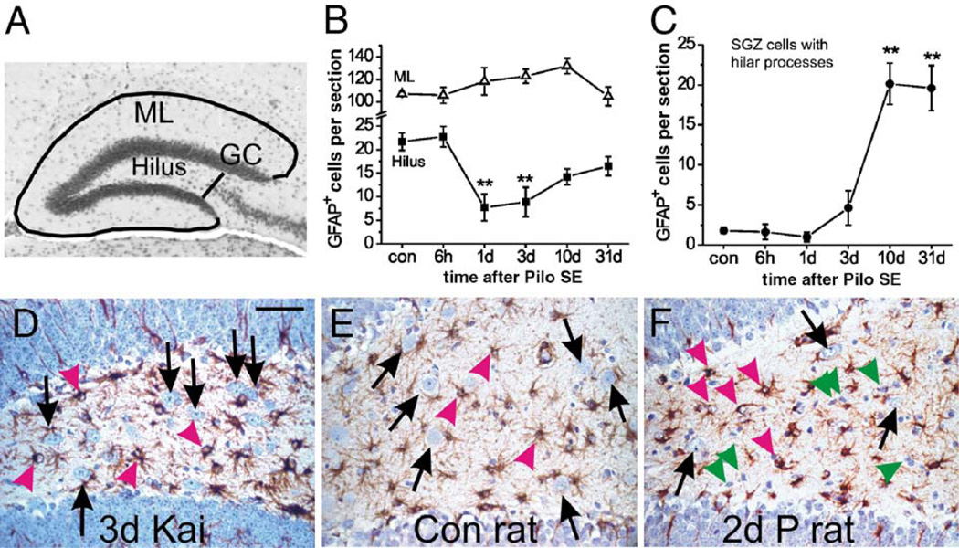
Fig. 2.
Selective astrocyte death in the dentate hilus after pilocarpine-induced SE is dependent on species and method to induce SE. (A) The micrograph shows a typical dorsal dentate gyrus section with the sampled areas outlined. The hilar area is between the two blades containing the dentate granule cells and the black line perpendicular to the end of the CA3 pyramidal cell layer. The molecular layer (ML) is found outside the granule cell layer (GC) and bordered by the black lines. The average cell numbers and standard error of healthy GFAP-positive cells per 8 µm dorsal hippocampal section are shown at different times after pilocarpine-induced SE (n = 4 – 5 CF1 mice) for the dentate molecular layer (B, triangles), the hilus (B, squares) and subgranular astrocytes that show hilar processes (C). The number of healthy GFAP-expressing cells significantly declined 1 and 3 days after SE in the hilus, but not the dentate molecular layer (one-way ANOVA followed by a post hoc Dunnett test). The number of subgranular cells with hilar processes significantly increased ten and 31 days after SE (one-way ANOVA followed by a post hoc Dunnett test). All comparisons are relative to control mice. **P < 0.01. (D–F) Representative GFAP immunostainings (brown) of the hilus are shown from a CF1 mouse 3 days after about 4 h SE induced by repeated kainate injections (D), a control rat (E) and a rat 2 days after 2.5 h SE induced by pilocarpine (F). Note that GFAP-expressing cells appear healthy (pink arrowheads) in all three sections. Hilar neurons (black arrows) are reduced in number after SE in the rat (F), but not in the mouse after kainate-induced SE. Some shrunken GFAP-negative cells, most likely dying neurons, are pointed out by green arrowheads. Sections are counterstained with hematoxylin and the scale bar is 50 µm. (For interpretation of the references to colour in this figure legend, the reader is referred to the web version of this article.)

