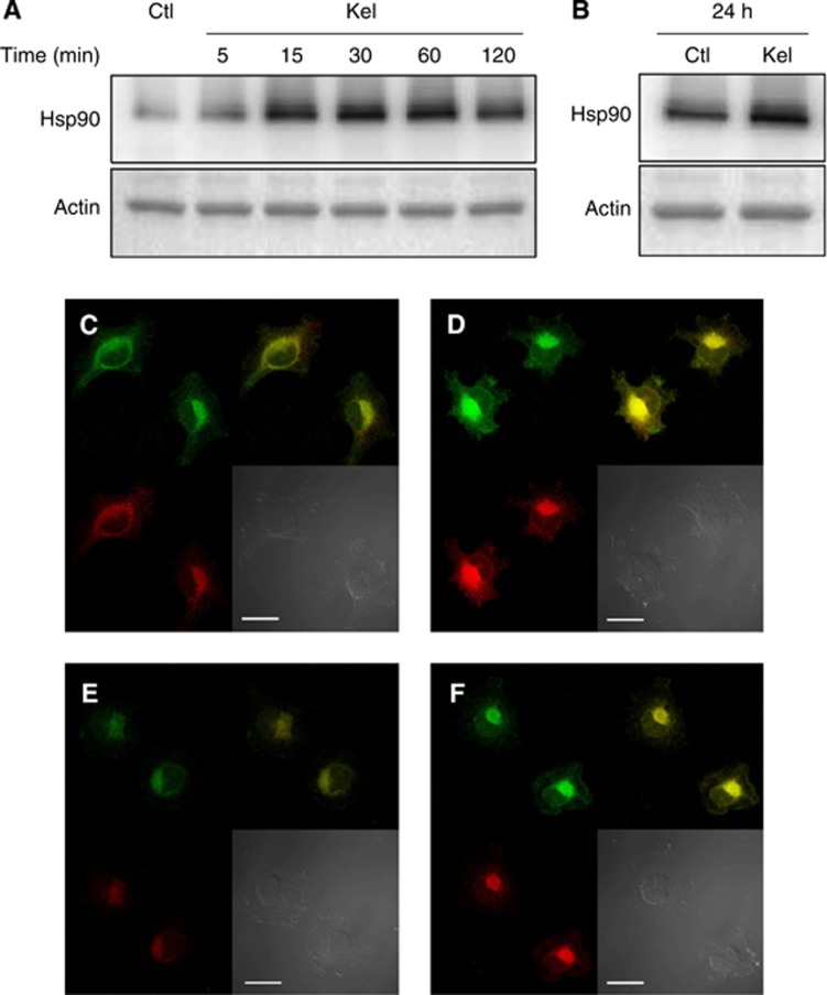
Figure 2.
Evaluation of the intracellular Hsp90, MMP-2 and uPA expression in HT-1080 cells. (A) Western blotting analysis of intracellular Hsp90 using a rabbit anti-Hsp90 monoclonal antibody and probing with anti-actin antibody were carried out as described in Materials and Methods section. (B) Western blotting analysis of intracellular Hsp90 at 24 h using a rabbit anti-Hsp90 monoclonal antibody and probing with anti-actin. (C–F) Localisation of Hsp90, pro-MMP-2 and uPA in HT-1080 cells in the presence (D, F) (Kel 50 μg ml−1) or absence (C, E) (control) of EDPs. Cells were cultured on glass slides, fixed with paraformaldehyde and labelled with an anti-Hsp90 (green), anti-pro-MMP-2 form (red) (C, D), anti-uPA (red) (E, F). Yellow staining corresponds to areas where Hsp90, pro-MMP-2 or uPA colocalised. Scale bar: 10 μm.

