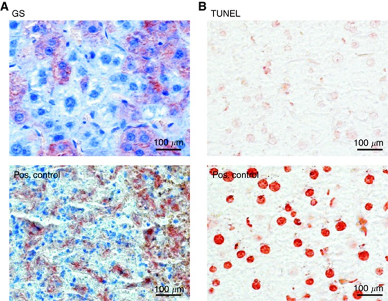
Figure 3.
Absence of necrosis and inflammation from Ctnnb1 KO livers. (A) Intact GS-stained tumour tissue taken from a Ctnnb1 KO animal versus positive ‘Pos.' control showing tissue from a highly necrotic tumour infiltrated by immune cells and with remnants of former GS-positive hepatoma cells (sample taken from a previous study by Singh et al (2013)). (B) Tumour tissue from A stained by TUNEL technology reveals the absence of apoptotic cell nuclei after KO of Ctnnb1. Positive control was pre-treated with benzonase nuclease to generate free DNA ends. Liver was taken from a Cre-positive, Ctnnb1 KO animal 3 weeks after tamoxifen treatment.

